Fantastic Poses and Can We Even Find Them? - What we learned about PXR structure prediction in our recent challenge
Co-folding has taken over protein-ligand structure prediction, but can it find the right pose? Key learnings from PXR structure prediction in our recent blind challenge.

Authors: Maria Castellanos, Jon Swain and Hugo MacDermott-Opeskin
DOI: https://doi.org/10.5281/zenodo.23408091
In Part I of our PXR Challenge series, we broke down the results of the Activity Track, analyzing how the top teams tackled the very difficult task of predicting PXR induction on a test set packed with activity cliffs. Now, we turn to the Structure Track.
Unintended PXR (pregnane X receptor) agonism is a major liability in drug discovery, as it triggers upregulation of key CYP enzymes such as CYP3A4. Because PXR features an exceptionally flexible, promiscuous binding pocket, accurately modeling its protein-ligand interactions presents a formidable challenge. Across nearly 100 competing teams, the top submissions revealed crucial lessons about co-folding architectures, fine-tuning, and the continued dominance of physics-based force fields for pose ranking. Let’s dive in!
Benchmarking protein-ligand structure prediction against 184 new crystal structures
PXR features an unusually large, flexible binding pocket (or is thought to be flexible; stay tuned for an upcoming blog post on this topic!), enabling it to act as a master xenobiotic sensor by accommodating diverse ligands across a wide range of shapes and sizes [1]. However, this extreme structural flexibility makes PXR a notoriously difficult target for structure-based computational methods [2]. In particular, the binding site is likely capable of binding small molecules in multiple orientations and/or fit multiple fragments at the same time. Because of this, ligand-based approaches such as QSAR, pharmacophore modeling, and machine learning have traditionally achieved somewhat better success in predicting PXR agonism than structure-based methods [1].
To establish a rigorous benchmark for state-of-the-art protein-ligand structure prediction on key ADMET enzymes, the Fraser lab solved a dataset of 184 X-ray crystallography structures of PXR (the largest publicly available PXR complex dataset to date!) bound to various fragments and drug-like compounds from our screens at Octant.
This demanding target challenged participants. We saw a broad range of creative workflows that combined state-of-the-art co-folding models, physics- and ML-based scoring functions, and novel sampling techniques. Below, we break down these strategies and summarize what they reveal about the current state of protein-ligand modeling.
Summary of strategies employed by Tier 1 teams
In this Structure Track, we also observed very close performance between teams. A total of 10 submissions demonstrated performance that was not statistically distinct from the #1-ranked model, comprising Tier 1. The interactive table below summarizes the core methodologies deployed by Tier 1 teams across key technical parameters:
- How many and which co-folding models were used
- Whether Multiple Sequence Alignment (MSA) was used as a starting point
- If Docking methods were used
- Whether templates or external data were used to improve the folding
- If Physics-based approaches were used in the strategy
- Whether fine-tuning of the co-folding models was attempted
- Strategies used for Pose Ranking
- Validation methods used
Table 1. Summary of methods participants used in the Structure Track. Hover over symbols and numbers to see details and click abbreviated column headers to expand them. A ✓ means the method or strategy was used in the final submission, ✗ means it was tried but not used in the final submission, and — means it was not used or not mentioned in the report. For the MSA column: ✓ custom/fixed MSA, ◐ model-default MSA, ✗ no MSA.
Co-folding universally outperforms fixed-receptor docking
Co-folding was the clear method of choice among Tier 1 participants. Most top teams combined multiple co-folding architectures into unified pipelines, noting that this produced better results. Rather than relying on a single network, participants commonly used co-folding algorithms to generate broad ensembles of candidate poses, which were then filtered and ranked using custom scoring pipelines.
On the other hand, traditional fixed-receptor methods took a back seat. Because PXR's binding pocket is thought to undergo substantial induced-fit rearrangements [1], rigid or semi-flexible docking methods performed poorly relative to deep co-folding models capable of co-optimizing protein side-chain/backbone conformations and ligand geometry. Classical docking was used by only two Tier 1 teams: TangerineTrees (Glide/ICM-Pro) and dargason (GNINA). In both cases, it served as a conditional fallback or anchor-specific sanity check rather than the primary pose generator.
ESMFold2 is crowned the new co-folding queen
Generally, the method of choice across top-performing teams was to combine multiple co-folding models to sample a diverse pool of candidate poses. Among them, ESMFold2, a structure prediction model built on the ESMC protein language model (pLM), was one of the most widely used by top teams, and some teams even reported that it outperformed AF3-style co-folding models such as Boltz-2 and OpenFold3. For example, willvith (#1 and #2) ran ESMFold2 with a fixed Multiple Sequence Alignment (MSA), both fine-tuned and as the base model, while other teams, such as Radi (#3), relied instead on AlphaFold3 as their main co-folding model.
One of the most surprising findings was the outstanding performance of single-sequence ESMFold2. Systematic benchmarking by dargason (#9), who could not run the MSA mode due to memory constraints, found that this model was "head-and-shoulders above everything else" at generating accurate starting candidate poses. This result is particularly striking given that single-sequence ESMFold2 uses no explicit MSAs and is less memory-intensive.
We speculate that ESMFold2’s advantage lies in its pLM core [3]. Pretraining on a massive database of ~2.8 billion metagenomic sequences may enable the model to internalize much of the evolutionary signal that an MSA would otherwise provide. This would explain why the single-sequence mode remains competitive with other MSA-based models, while the ESMFold2 paper reports that accuracy improves further when MSAs are supplied [3], suggesting the two sources of information (namely, the ESMC pLM and MSAs) are complementary. PXR’s large, highly flexible, and promiscuous binding pocket makes it a challenging target, so a strong learned prior may be especially useful here. However, differences in training data and cutoff dates between models could also contribute to the difference in performance. In an upcoming blog post, we will dive deeper into the performance of each co-folding model and their confidence scores on the PXR data.
Another popular model was Protenix-v2 [4], an open-source AlphaFold3-style model, used by teams such as willvith, xX-its-amit-Xx, and NamuICT. Interestingly, xX-its-amit-Xx and NamuICT used this method as a “tail-rescue” model for hard-to-predict outliers. After selecting poses from their primary co-folders, they identified the 8 ligands with the lowest confidence (tuned on the leaderboard), for which no model was confident, and replaced each with the highest-ranked Protenix-v2 pose from pools of 25 and 100 samples per ligand, respectively. This strategy paired broad cross-model generation across all ligands with deeper, single-model sampling on a smaller, more challenging subset.
Physics-based methods: “You aren’t getting rid of me just yet”
Co-folding models excel at quickly generating candidate poses, but relying on their internal confidence scores (such as ipTM) for pose ranking can be unreliable. In practice, ipTM scores often correlate poorly with actual pose accuracy because they can be fooled by unphysical geometries. Furthermore, ML confidence estimators are naturally biased toward their training-set distributions, making them ill-suited for evaluating novel or unusual ligand conformations.
To overcome this limitation, top-performing teams pooled a larger volume of candidate poses and went beyond default confidence metrics, often re-ranking the candidates using dedicated scoring methods. Approaches split roughly into two categories: re-ranking with physics-based scoring, and combining confidence signals across models. Some of the physics-based approaches included:
- Using ML Force Fields: The top-performing team (willvith and e-phy) relied on machine learning interatomic potentials (MLIPs) as their core ranking signal. They calculated explicit binding interaction energies ($\Delta E = E_{\text{bound}} - E_{\text{protein}} - E_{\text{ligand}}$) using tools like Egret-1 and OrbMol-v2, paired with PoseBusters validity checks.
- Bypassing Confidence Metrics Entirely: Demonstrating the power of explicit physics, team ver228 reached Tier 1 using OrbMol-v2 energy alone, completely ignoring co-folding confidence scores like ipTM.
While co-folding models do well at sampling potential structural states, grounding final pose selection in explicit force-field interaction energies proved effective for identifying true binding poses (with willvith taking the #1 spot by combining physics scores with ipTM, and ver228 reaching Tier 1 with interaction energies alone). That being said, physics-based strategies didn’t always lead to a win: dargason found MM-GBSA unhelpful, and mittface's MMFF-strain selectors all scored below confidence-based selection.
An alternative strategy that preserved per-model confidence was to combine scores from different models. For example, xX-its-amit-Xx and NamuICT calculated the z-scores of each model’s native confidence signal across all 184 ligands, then picked the model with the highest z-score per ligand. This strategy allowed them to normalize contributions from different scores and increase coverage for molecules where any single model failed.
You can use fine-tuning to improve performance, but carefully
Fine-tuning co-folding models on target-specific data has become an increasingly common strategy, but this challenge is a reminder that it’s only as good as the data it's trained on: helpful when the fine-tuning set resembles the distribution of the model’s test set, but counterproductive when it doesn’t.
Few teams attempted to fine-tune co-folding models, and the results were mixed. willvith (#1) fine-tuned ESMFold2 on a curated set of 64 PXR crystal structures, paired with synthetic distillation data from ChEMBL, PubChem, and Enamine, and included both the base and fine-tuned models in their ensemble. By contrast, bear (#4) tried to fine-tune OpenFold3, which slightly improved cross-validation on Butina-clustered re-refined PXR structures, but hurt performance on the live leaderboard. In a related failed attempt, dargason (#9) trained a pose-picking (AI-assisted) function on co-folded, re-refined structures, which also failed to generalize to the leaderboard. Meanwhile, several Tier 1 teams (e.g., ver228, NamuICT) used models only at inference, with no fine-tuning.
A likely culprit is a train/test distribution mismatch: the re-refined PXR structures are a heterogeneous mix, including fragments, drug-like compounds and metal-containing complexes, while the test set (particularly the phase 1 set) was more fragment-heavy. Consistent with this, bear noted that improvements on the re-refined set “never transferred to the leaderboard”, and wondered whether fine-tuning OpenFold3 would have worked better for ligands closer to the re-refined structures. A similar mismatch also affected pose selection for mittface, who found that every test-set fragment had Tanimoto similarity below 0.3 to every known PXR holo ligand. As a result, signals that were tuned to drug-like structures (e.g., a CNN scorer, a PDBbind-trained model, and a ChEMBL-derived head) inverted on the test-set fragments. The takeaway is that fine-tuning is only as good as how well its training data reflects the target chemistry; in this case, neither the re-refined set nor external drug-like datasets were a good proxy for the test set, for fine-tuning and pose selection alike.
Looks like there wasn’t much external data to use this time
Similar to the Activity Track, proprietary and external experimental datasets were virtually nonexistent for this target. Instead, performance differences came down to co-folding model selection and scoring/ranking functions to select from ML-sampled poses (as mentioned in the previous sections). Another strategy that set top participants apart was how best to leverage the limited public data available for either fine-tuning or template selection for MSA.
Rather than relying on automated searches over large database dumps, top performers found that small, carefully hand-picked template sets performed best. For example, Radi embedded a precomputed MSA and only four fixed PDB templates (6S41, 7CHG, 5A86, 2O9I) directly in the AlphaFold3 input, disabling its automated template pipeline so no per-run search was performed. TangerineTrees benchmarked three distinct templates and identified 9FZJ as a consistent structural guide, reaching an LDDT-PLI of 0.529, better than both AlphaFold3 with automated template search (0.497) and plain AlphaFold3 with no template at all (0.506).
On the other hand, bear observed that providing a large, unfiltered pool of templates via Boltz-2 provided little performance improvement and introduced significant selection-overfitting risk.
Where did everyone fail?
As with the Activity Track, we investigated performance patterns on the compounds that participants universally struggled with or easily solved.
For the Structure Track, the primary metric was LDDT-PLI (Local Distance Difference Test for Protein-Ligand Interactions); a superposition-free metric that evaluates local atomic distance differences. Our secondary metric was BiSyRMSD (Binding-Site Superposed, Symmetry-Corrected Pose Root-Mean-Square Deviation), which superimposes the binding site and computes the symmetry-corrected ligand RMSD relative to the crystal structure.
Some compounds were universally harder or easier to predict
To determine whether performance trends correlated with compound identity, we plotted the Tier-1 average LDDT-PLI and BiSyRMSD scores per compound (Figure 1), ordered by increasing LDDT-PLI (where higher LDDT-PLI and lower BiSyRMSD indicate better prediction quality). Data points are color-coded by their originating library: fragments (orange), analogues (blue), or diversity compounds (green).
Figure 1: Distribution of LDDT-PLI (top) and BiSyRMSD (bottom) per compound across Tier-1 participants (ordered by increasing LDDT-PLI). Data points are colored by each compound-originating library: fragments (orange), analogues (blue), or diversity (green). Interactive view allows hovering over data points to display compound IDs.
Across Tier-1 submissions, predictions showed remarkable consensus at the extremes: the hardest compounds were universally difficult for all teams, and the easiest were universally well predicted. We also noted subtle patterns based on library type. While a few fragments proved remarkably challenging, fragment-like compounds generally showed higher prediction accuracy across both metrics. Conversely, no clear performance patterns emerged between the diversity and analogue libraries. To understand the drivers behind these trends, we tested several hypotheses.
Are larger compounds harder to predict?
Naturally, our first hypothesis was that performance differences between libraries stemmed primarily from molecular size. Namely, that fragments are generally smaller than lead-like compounds. In Figure 2, we visualized Tier-1 performance using box plots of per-compound scores binned by molecular weight in 50 Da increments (with individual fragment and non-fragment compounds overlaid). We observed a moderate negative correlation between MW and LDDT-PLI ($r = -0.33$). While not all compounds followed this trend (with a few fragments being harder to predict), a correlation still seems to exist.
Interestingly, the correlation weakens when measured by BiSyRMSD ($r = 0.25$). Because LDDT-PLI evaluates pairwise interatomic contacts, larger ligands with more atoms and contact pairs offer more chances for small local geometry errors to accumulate and lower the final score. BiSyRMSD is less sensitive to the total atomic count, so it shows a milder size dependence.
To look beyond simple metrics, we examined whether chemical complexity explains this size trend.
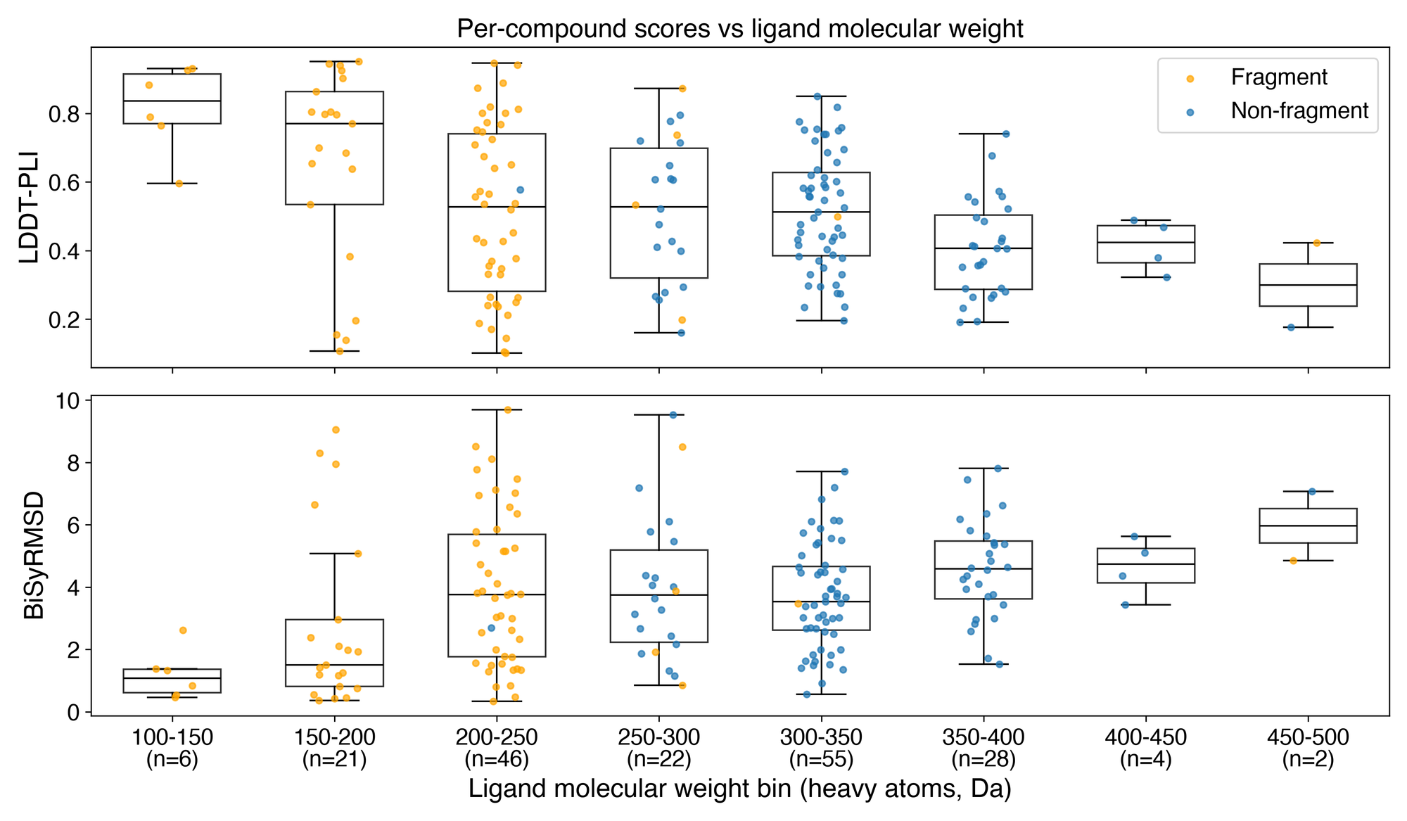
Figure 2. Distribution Tier-1 performance as a function of ligand molecular weight (binned every 50 Da). Box plots show the score distribution (median and IQR) for LDDT-PLI (top) and BiSyRMSD (bottom), with individual compounds overlaid as jittered points colored by library type (orange = fragment, blue = non-fragment). A negative correlation with molecular weight is observed for LDDT-PLI ($r = -0.33$), alongside a weaker positive correlation for BiSyRMSD ($r = 0.25$).
No clear link between ligand chemistry and its pose accuracy
We explored three potential mechanisms that could drive lower prediction accuracy in higher molecular weight compounds:
-
Combinatorial search difficulty. Traditional docking struggles with flexible molecules because the sampling space grows exponentially with the number of rotatable bonds [6]. In co-folding models, high flexibility requires sampling a far broader pose landscape, increasing the likelihood that a ranking function selects an incorrect pose. However, when evaluating performance against rotatable bond count (Figure 3, top), we found that while rigid molecules (0–1 rotatable bonds) were indeed easy to predict, performance plateaued and showed no correlation for molecules with $\geq 2$ rotatable bonds.
-
Presence of specific large functional groups: Another hypothesis is that certain large functional groups might drive complex protein-ligand interactions or induce binding-pocket shifts that confound co-folding models. We identified the most common functional group motifs among the 20 hardest-to-predict compounds and evaluated their distributions (Figure 3, bottom). The presence of specific functional groups showed no clear relationship with prediction error.
-
Larger ligands promote more induced-fit rearrangement. PXR possesses a notoriously flexible binding pocket that can expand by more than $400\ \text{Å}^3$ depending on the bound ligand [1]. Because models like AlphaFold3 are known to struggle with complexes requiring substantial conformational shifts ($>5\ \text{Å}$ RMSD), larger ligands might promote larger induced-fit rearrangements. However, when we calculated the binding-site backbone RMSD for the 20 hardest compounds across the top three teams, all predictions stayed under $1\ \text{Å}$, which rules induced fit out as the cause of the MW dependence. It is worth noting that the reference structures are themselves highly similar to one another (with pairwise binding-site RMSD ranging between $0.09\text{–}1.47\ \text{Å}$, and a median of $0.34\ \text{Å}$), so this crystal system samples only a narrow fraction of PXR’s conformational range, and these predictions are no evidence that co-folding models capture PXR’s full flexibility.
As it seems, there is no chemically grounded explanation for the negative correlation between LDDT-PLI performance and MW, so we cannot say for certain that fragments or smaller ligands are easier to predict than drug-like or larger compounds. The core limitation must rest in the co-folding models' inability to place ligands accurately.
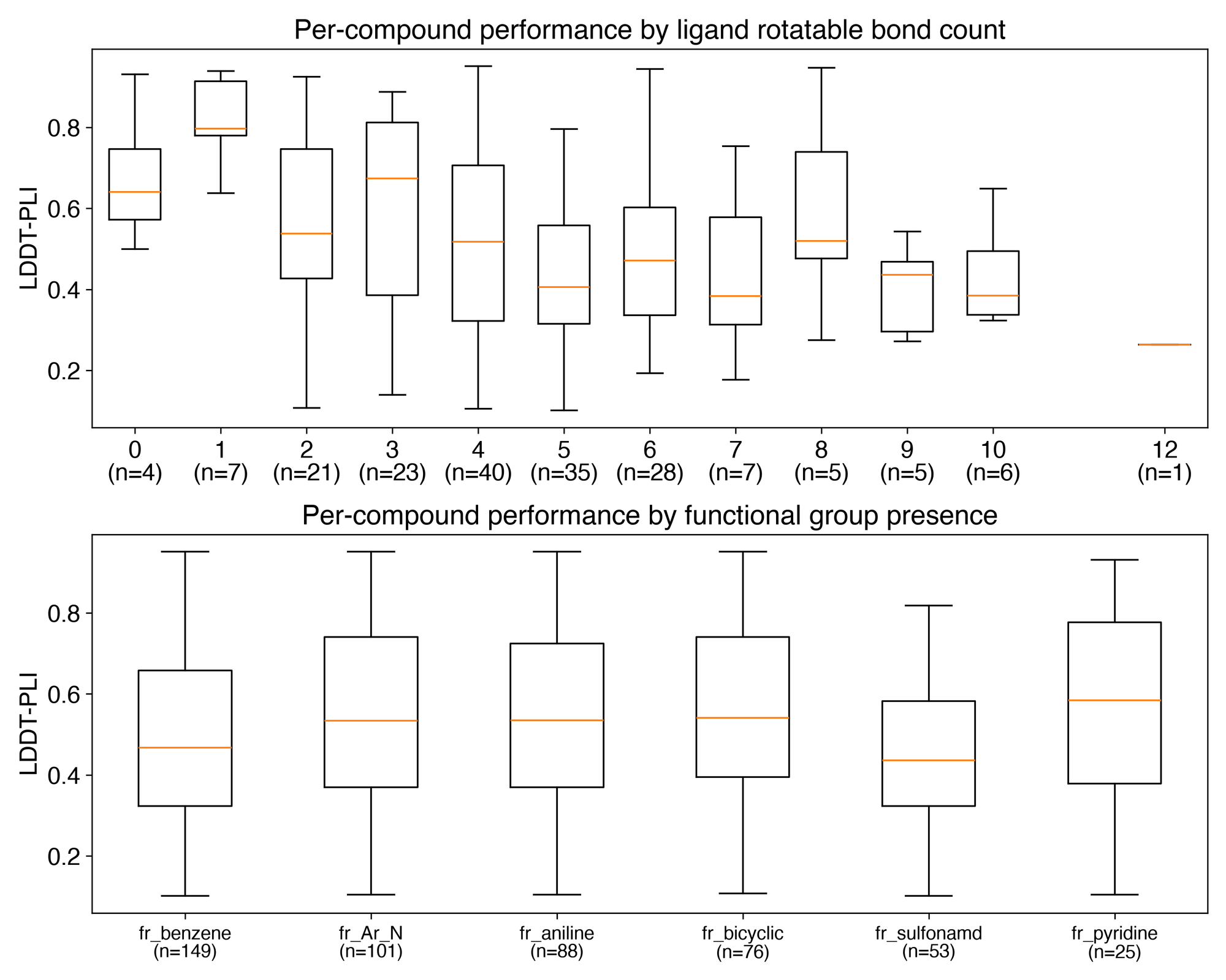
Figure 3. Distribution of Tier-1 LDDT-PLI performance vs. ligand rotatable bond count (top) and presence of key functional groups (bottom). Sample counts per category are shown in parentheses.
Co-folding ain’t beating the “memorization” allegations
Our findings point to a central challenge in structural AI: co-folding architectures struggle to generalize to protein-ligand conformations that are underrepresented in, or absent from, their training distributions [5].
PXR presents a twofold challenge for data-driven models, with each aspect acting at different stages of the prediction process. First, at generation: PXR crystal structures in the PDB are (besides being sparse) dominated by specific chemotypes, biasing models toward memorized poses. Novel ligands outside this distribution are prone to severe mispredictions. Second, at selection: PXR’s large, flexible, and largely hydrophobic pocket means fragments make few specific contacts, so several placements are geometrically plausible (a point raised by Radi in their report). As we demonstrated in the previous section, the models get the pocket right; what they can’t do is identify which ligand pose within it is correct, and the confidence metrics used to rank candidates also discriminate poorly.
It is hard to say which of these (e.g., models failing to generate the correct pose or ranking strategies failing to select the correctly predicted poses) hurt leaderboard performance the most based on challenge submissions alone. Either way, it seems that our current methods are not yet capable of reliably predicting poses outside their training distribution.
Could pose predictions in this challenge translate to correct SAR?
Finally, we evaluated whether predicted poses capture true protein-ligand interaction profiles. Protein-ligand interactions are the foundation for using predicted structures in structure-based activity modeling and SAR expansion.
In Figure 4, we compare the protein-ligand interaction fingerprints of two of the hard-to-predict (left) and two of the easy-to-predict compounds (right), comparing ground-truth crystal structures against predictions from the top three teams (willvith, Radi, and bear). The interactions are presented as a barcode, as calculated with ProLIF, indicating whether the residues (y-axis) form Hydrophobic interactions, Hydrogen bonds as either acceptors (HBAcceptor) or donors (HBDonor), and Van der Waals (VdW) contacts. The poses of one hard-to-predict (x03273-1) and one easy-to-predict (x00409-1) structure are shown on the right. The LDDT-PLI and BiSyRMSD of each prediction are shown in the bar plot at the bottom.
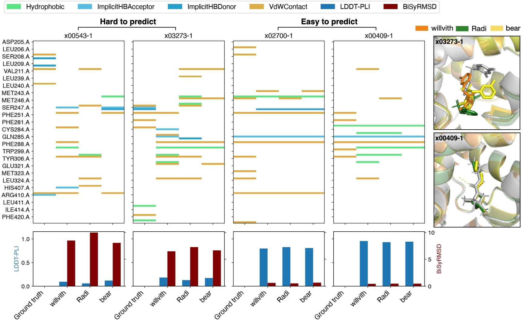
Figure 4. Interaction fingerprint barcode (as calculated with ProLIF) for two of the compounds that were incorrectly predicted by most participants (x00543-1 and x03273-1), and two of the compounds that were universally easier to predict (x02700-1 and x00409-1), for the ground truth compared to the predictions of the top 3 teams: willvith, Radi, and bear. While the pose seems to be accurately predicted (right panel), the protein-ligand interactions don’t completely match the ground truth.
The main takeaway is that, for difficult compounds, the models completely failed to capture the true interaction fingerprints. Hydrogen bonds, salt bridges, and hydrophobic contacts were severely scrambled relative to ground truth. As shown in the structural overlays, predicted poses adopt fundamentally incorrect binding orientations. While in easy-to-predict compounds, core scaffolds were oriented correctly, and key backbone-anchored interactions were preserved, subtle differences in substituent positioning led to discrepancies in hydrophobic and Van der Waals contacts. Note that these barcodes depend on ProLIF’s interaction definitions and distance and angle cutoffs, such that a pose sitting near a threshold may gain or lose an interaction for a sub-ångström coordinate shift. This is more likely to affect the subtle differences seen in the easy-to-predict compounds than the larger mismatches in the hard cases.
Because LDDT-PLI weights core scaffold contacts heavily, minor substituent shifts do not heavily penalize the overall score, even though those exact substituent interactions often dictate SAR. For difficult compounds, where key interactions are missed entirely, structure-based activity modeling would be completely unviable.
While top co-folding pipelines can reliably place core scaffolds for compounds that are easier to predict, they still fail to capture fine-grained substituent interactions. It remains to be seen if these differences could lead to differences in the SAR and in potential structure-based activity predictions.
Who You Gonna Call? PoseBusters!
While AI co-folding models clearly led the pack over traditional physics-based docking in this challenge, headline performance metrics like BiSyRMSD and LDDT-PLI don’t always tell the whole story. A pose can achieve an outstanding BiSyRMSD or contact score while simultaneously violating basic laws of physical chemistry.
This was demonstrated by Buttenschoen, Morris, and Deane in their 2024 PoseBusters paper. [7] They showed that applying strict physical plausibility filters (the "PB-valid" benchmark) substantially degrades the reported success rates of deep-learning-based structure prediction tools.
With that in mind, we ran our top-tier submitted structure predictions through the PoseBusters suite.
It’s important to note that PoseBusters validity was not included in our official scoring criteria for this round. Had a validity filter been an explicit requirement, participants likely would have adapted their workflows; for instance, by adding constrained energy minimizations or physics-based relaxation steps. This analysis is purely post-hoc, though we intend to incorporate validity checks directly into future challenge leaderboards.
Of the 1,840 submitted poses analyzed in the top tier (10 submissions of 184 structures each), not a single submission was entirely PB-valid across all targets. Individual submission validity ranged between 131 and 181 out of 184 structures. This is despite several teams screening for physical plausibility themselves. For example, willvith folded the PoseBusters score directly into their ranking function, weighting it heavily against predicted interaction energy; dargason filtered on PoseBusters and then applied a restrained minimization to relax residual clashes and strain, while bear and ver228 filtered on chirality errors and atomic forces, respectively. That even these submissions contained invalid poses suggests the problem is not simply one of post-processing.
Across the dataset, we observed a small number of severe geometric anomalies: non-flat aromatic rings (2), high internal energy (3), and volume overlap with the protein (3). More commonly, entries failed due to non-aromatic rings being predicted as planar (21) and internal ligand steric clashes (39).
The instances of non-aromatic rings being predicted as planar offer an interesting insight into how coordinate-generating deep-learning models operate. Because many co-folding architectures predict raw 3D Cartesian coordinates directly, relying on interatomic distance or template priors rather than explicit valence bond physics for bond orders, they can inadvertently flatten saturated or partially saturated rings as if they were planar aromatics. The most common offending molecules (Figure 5) all contain a cyclohexyl group, which deep learning methods may have predicted as a benzene ring.
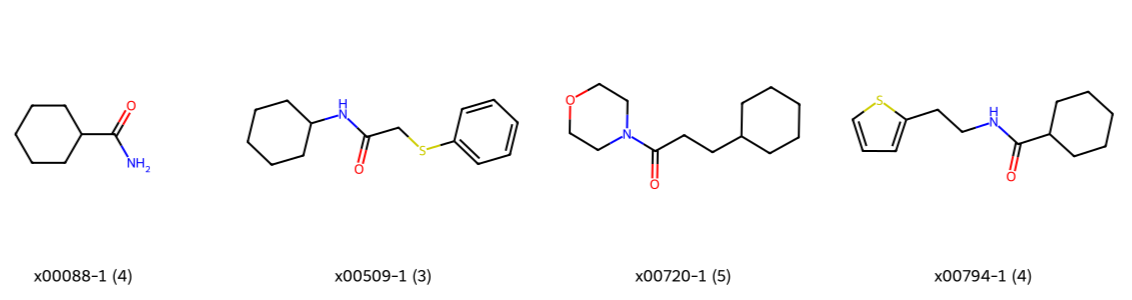
Figure 5. Structures of ligands commonly predicted with planar non-aromatic rings. Numbers in brackets indicate prevalence across the top tier of submissions (n=10 entries). All flagged poses contain saturated cyclohexyl rings collapsed into planar geometries.
Among the predictions with internal ligand steric clashes, we observed a consistent pattern: all offending structures shared a conserved urea core directly bonded to an aromatic ring (Figure 6). In each case, the steric clash was between a urea nitrogen and atoms on the ring on the far side of the urea, specifically the ring heteroatom and the ring carbon bearing the urea. In fact, one ligand (x03259-1) was predicted to contain internal ligand steric clashes by 9 of the top 10 participants. The urea nitrogen beside the cyclobutyl group clashed with the carbon and first nitrogen atoms of the triazole ring. For five of the seven ligands with internal steric clashes, the experimental reference structure also fails the check at the same atoms, indicating that either PoseBusters' distance‑geometry bounds are too strict, or the uncertainty in the crystal structures needs to be accounted for if using PoseBusters as part of a scoring pipeline.
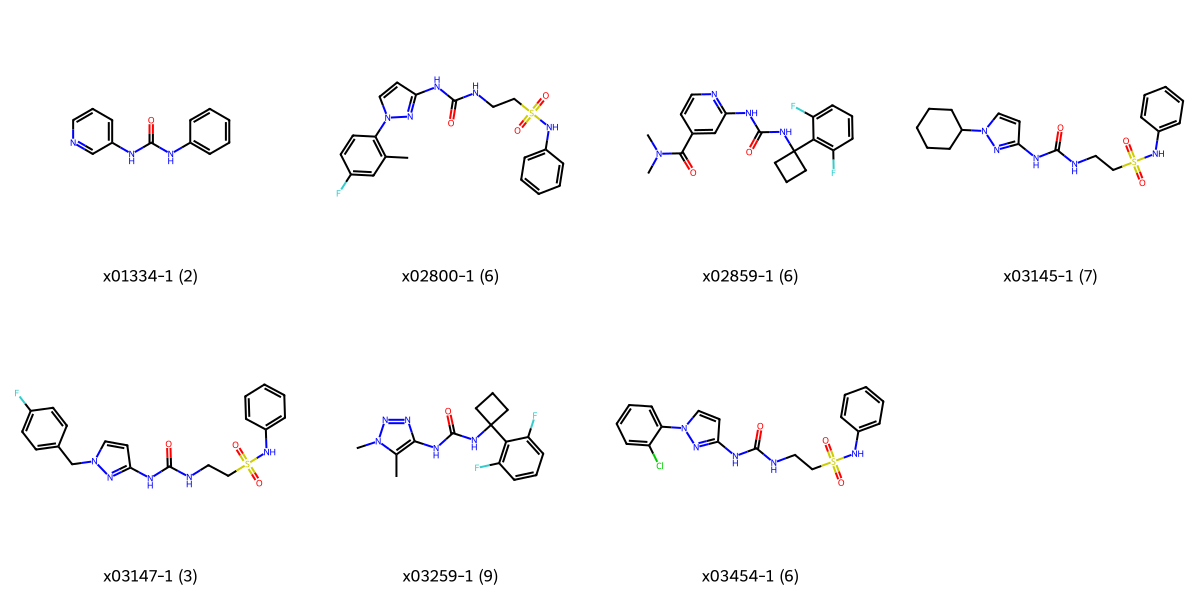
Figure 6. Structures of ligands containing internal ligand steric clashes. Numbers in brackets denote failure count across the top tier (n=10 entries), highlighting a persistent vulnerability in maintaining valid urea backbone geometries.
By far the most prevalent issue, however, was minimum distance violations to the protein (148 cases). This highlights a fundamental limitation of standard evaluation metrics. Because tight binding pockets naturally involve short contact distances, an unconstrained generative model predicting poses near the binding threshold will inevitably produce poses that violate physical constraints. Crucially, metrics like BiSyRMSD and LDDT-PLI reward ligand overlap and contact recovery without heavily penalising unphysical clashes. Across the 10 top-tier entries, the per-compound prevalence of minimum-distance violations ranged from 0 to 5. Interestingly, no correlation was observed between molecular weight and clash frequency; fragments were just as prone to clashes as larger molecules. Minimum distance violations to the protein were also flagged by PoseBusters in 12 of the ground-truth structures, again showing that incorporating PoseBusters into a scoring pipeline will require more than just rejecting entries that are not PB-valid.
PoseBusters proved to be an invaluable diagnostic for highlighting these subtle failure modes, and physical validity filters will be an integral part of how we evaluate and score entries in future blind challenges.
Conclusions
It is encouraging that teams are working around the limitations of co-folding confidence scores by using these models for pose sampling, and leveraging physics-based approaches (which we understand well) to choose among the sampled pool of candidates. The right poses are often in the ensemble; it's just a matter of pulling out the right one.
However, applying co-folding models to out-of-distribution ligands still needs work, even with the strategies participants adopted. It is hard to tell whether the models fail to predict molecules unlike those in their training data, whether the selection strategies have room for improvement, or both.
Also encouraging is the strong performance of ESMFold2, a model built on a protein language model. It seems pre-training on large, diverse sequence databases may be an important ingredient for accurate structure prediction on difficult ADMET targets like PXR, though other factors, such as differences in training data between models, could also play a role.
Finally, fine-tuning showed that data quality matters as much as quantity: it helped when the data resembled the model’s original distribution but failed when it did not match the fragment-heavy test set. We still have a long way to go for protein-ligand structure prediction, but rich datasets on targets that are traditionally hard to predict (as well as critical for ADMET) will allow current models to improve and foster more creative approaches to protein-ligand prediction.
Acknowledgements
We would like to acknowledge the hard work of all the experimentalists at Octant and UCSF for making this challenge possible. In particular, we would like to thank Sam Sabaat, Scott Simpkins, Yuning Shen, Bryan Jiang, Henry Chan, Jeff Tang, Ayesha Ghazali, Theo Tarver, Steven Edgar, Dominic Ky, and many others from Octant, and Galen Correy, Nikhil Gupta, and others from the Fraser lab at UCSF. We also thank the beamline staff at NSLS-II.
We would like to thank our funders for their support of OpenADMET, in particular ARPA-H, Radial (part of the Astera Institute (https://ror.org/00ydx1s47)), Schrödinger Inc., the Gates Foundation and the OpenAI Foundation. We would also like to thank our partners Enamine, HuggingFace, OpenEye, CDD Vault, Discovery Life Sciences, and the beamline staff at NSLS-II for their support.
This work is supported by the Advanced Research Projects Agency for Health (ARPA-H) under AVOID-OME, and Award Number 1AY1AX000035. The contents are those of the authors. They may not reflect the policies of the Department of Health and Human Services or the U.S. government. The content is solely the responsibility of the authors and does not necessarily represent the official views of the Advanced Research Projects Agency for Health.
References
[1] Watkins, R. E., Wisely, G. B., Moore, L. B., Collins, J. L., Lambert, M. H., Williams, S. P., ... & Redinbo, M. R. (2001). The human nuclear xenobiotic receptor PXR: structural determinants of directed promiscuity. Science, 292(5525), 2329-2333.
[2] Ekins, S., Kortagere, S., Iyer, M., Reschly, E. J., Lill, M. A., Redinbo, M. R., & Krasowski, M. D. (2009). Challenges predicting ligand-receptor interactions of promiscuous proteins: the nuclear receptor PXR. PLoS computational biology, 5(12), e1000594.
[3] Candido, S., Hayes, T., Derry, A., Rao, R., Lin, Z., Verkuil, R., ... & Rives, A. (2026). Language modeling materializes a world model of protein biology. bioRxiv, 2026-06.
[4] ByteDance AML AI4Science Team, Chen, X., Zhang, Y., Lu, C., Ma, W., Guan, J., ... & Xiao, W. (2025). Protenix-advancing structure prediction through a comprehensive AlphaFold3 reproduction. BioRxiv, 2025-01.
[5] Škrinjar, P., Eberhardt, J., Studer, G., Tauriello, G., Schwede, T., & Durairaj, J. (2026). Evaluating generalization in protein–ligand cofolding methods. Nature Structural & Molecular Biology, 1-13.
[6] Berry, M., Fielding, B., & Gamieldien, J. (2015). Practical considerations in virtual screening and molecular docking. Emerging trends in computational biology, bioinformatics, and systems biology, 487.
[7] Buttenschoen, M., Morris, G. M., & Deane, C. M. (2024) PoseBusters: AI-based docking methods fail to generate physically valid poses or generalise to novel sequences. Chem. Sci. 15 (9): 3130–3139.