Analyzing CYP3A4 PDB structures
Looking at CYP3A4 X-ray structures from the PDB
Authors: Nikhil Gupta (UCSF)
DOI: 10.5281/zenodo.23871907
There are 109 ligand bound Cyp3A4 structures in the PDB, a rich source of information for model training. When building training/test sets for model training, we want to ensure that the data is of high quality. For structural data, this means ensuring that the structural model has a reasonably good agreement with the underlying experimental observables. Since we are particularly interested in the Cyp3A4 protein-ligand interactions, I will be focusing on how well the ligands of these structures fit into their experimental density.
Additionally, several ligands bind in allosteric sites or in not pharmacologically relevant conditions. These structures will not be useful for benchmarking performance and may confuse models during fine-tuning.
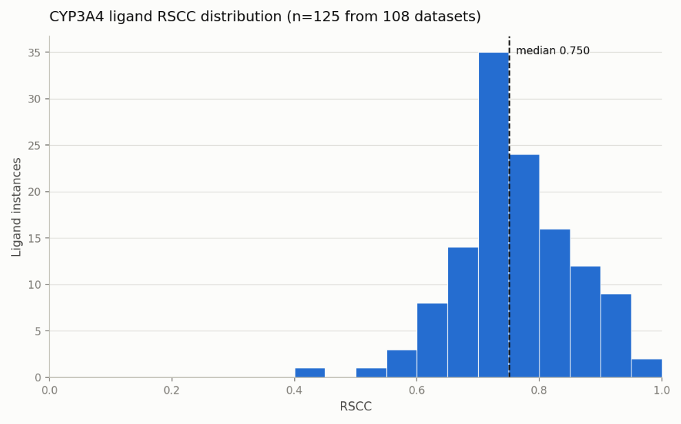
Fig 1: Real Space Correlation Coefficient (RSCC) of all ligands in the Cyp3A4 PDB dataset. What the score means is specific to each case but heuristics are approximately: an RSCC > 0.8 signifies a great fit, 0.7-0.8 an okay fit, 0.6-0.7 a questionable fit and < 0.6 a poor fit.
Most of the ligands in these datasets have good density fit, but there are several structures with ligands that I think are not sufficiently supported by their density. I will go through all of the PDBs that I think should be excluded when using the CYP3A4 PDB structures for training/evaluating models:
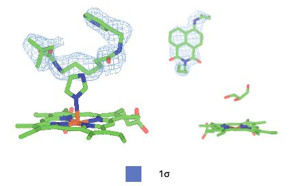
Fig 2: Structure 4D6Z (left) and 8DYC (right) both have good support, but have solvent molecules (imidazole for 4D6Z, glycerol for 8DYC) coordinating the heme and are not representative of the pharmacologically relevant state. Another structure, 4D75 has a structure with the same ligand as 4D6Z. In that structure, the ligand directly coordinates the heme.
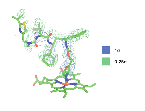
Fig 3: Structure 4I4G has low RSCC (0.66) and does not have strong density support visually. While the heme coordinating region of the ligand is fairly well resolved, much of the rest of the ligand is not. There are many other ritonavir-like structures that are better resolved.
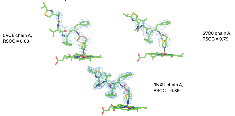
Fig 4: 5VCE, 5VC0, 3NXU, the ritonavir structures. All 3 structures have different poses and 3NXU has the best resolved ligand. I recommend eliminating 5VCE and 5VC0.
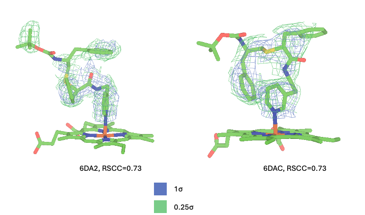
Figure 5: 6DA2 and 6DAC both have okay RSCCs, but visually have weak or no density for close to half of the ligand. While the ligands have okay density support, there are many other chemically similar ligands from the same paper with better density fit. This issue of ligand regions far away from the heme being poorly resolved has been something we see frequently in our cryo-em structures and prevents us from confidently modeling ligand poses for those datasets. Structures with many poorly resolved atoms can confuse structural AI models that treat coordinates as ground truth. This is an ongoing challenge for the field.
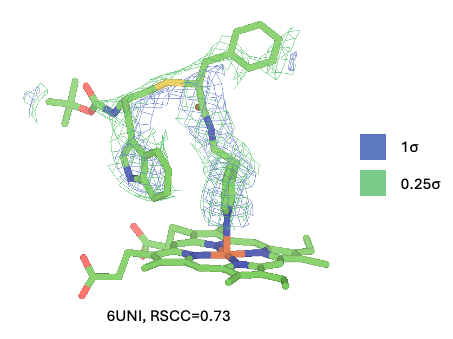
Figure 6: 6UNI has a decent RSCC, but visually fits its density quite poorly. The butyl and phenyl groups fit to blobs of density that are largely unconnected from the rest of the ligand density and don't fit those moieties well.
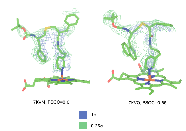
Figure 7: 7KVM and 7KVO both have very low RSCCs and visually have very weak fit, with significant regions of the molecules being very poorly resolved. 7KVO's other chain is even more poorly resolved, with an RSCC of 0.46.
Several of the structures from this paper have multiple chains: 7KVK, 7KVN, 7KVQ, and 7KVS. The ligands in these structures vary in quality between the two chains. For all 4 of these structures, I recommend considering only chain A.
Additionally, Structures 5A1P, 5A1R, and 1W0F all contain CYP3A4 bound to progesterone. These ligands have good support, but are bound in an allosteric site, and will not be useful for benchmarking/fine-tuning predictions that are focused on CYP3A4's primary binding site.
Recommendations
Here is a succinct table of my recommendations:
| Structure | Recommendation |
|---|---|
| 4D6Z | Exclude |
| 4I4G | Exclude |
| 5VCE | Exclude |
| 5VC0 | Exclude |
| 6DA2 | Exclude |
| 6DA3 | Exclude |
| 6UNI | Exclude |
| 7KVM | Exclude |
| 7KVO | Exclude |
| 5A1R | Exclude |
| 5A1P | Exclude |
| 1W0F | Exclude |
| 8DYC | Exclude |
| Structure | Recommendation |
|---|---|
| 7KVK | Take Chain A Only |
| 7KVN | Take Chain A Only |
| 7KVQ | Take Chain A Only |
| 7KVS | Take Chain A Only |
Funder Acknowledgement: We would like to thank our funders for their support of OpenADMET, in particular ARPA-H, Radial (part of the Astera Institute (https://ror.org/00ydx1s47)), Schrödinger Inc., the Gates Foundation and the OpenAI Foundation. We would also like to thank our partners Enamine, Hugging Face, OpenEye, CDD Vault, Discovery Life Sciences, and the beamline staff at NSLS-II for their support.
This work is supported by the Advanced Research Projects Agency for Health (ARPA-H) under AVOID-OME, and Award Number 1AY1AX000035. The contents are those of the authors. They may not reflect the policies of the Department of Health and Human Services or the U.S. government. The content is solely the responsibility of the authors and does not necessarily represent the official views of the Advanced Research Projects Agency for Health.